
Brain imaging of the cortex in ADHD: A coordinated analysis of large-scale clinical and population-based samples
Martine Hoogman, Ryan Muetzel, Joao P. Guimaraes, Elena Shumskaya, et al.
- Maarten Mennes, Marcel P. Zwiers, Neda Jahanshad, Gustavo Sudre, Jeanette Mostert, Thomas Wolfers, Eric A. Earl, Juan Carlos Soliva Vila, Yolanda Vives-Gilabert, Sabin Khadka, Stephanie E. Novotny, Catharina A. Hartman, Dirk J. Heslenfeld, Lizanne J.S. Schweren, Sara Ambrosino, Bob Oranje, Patrick de Zeeuw, Tiffany M. Chaim-Avancini, Pedro G. P. Rosa, Marcus V. Zanetti, Charles B. Malpas, Gregor Kohls, Georg G. von Polier, Jochen Seitz, Joseph Biederman, Alysa E. Doyle, Anders M. Dale, Theo G.M. van Erp, Jeffrey N. Epstein, Terry L. Jernigan, Ramona Baur-Streubel, Georg C. Ziegler, Kathrin C. Zierhut, Anouk Schrantee, Marie F. Høvik, Astri J. Lundervold, Clare Kelly, Hazel McCarthy, Norbert Skokauskas, Ruth L. O'Gorman Tuura, Anna Calvo, Sara Lera-Miguel, Rosa Nicolau, Kaylita C. Chantiluke, Anastasia Christakou, Alasdair Vance, Mara Cercignani, Matt C. Gabel, Philip Asherson, Sarah Baumeister, Daniel Brandeis, Sarah Hohmann, Ivanei E. Bramati, Fernanda Tovar-Moll, Andreas J. Fallgatter, Bernd Kardatzki, Lena Schwarz, Anatoly Anikin, Alexandr Baranov,Tinatin Gogberashvili, Dmitry Kapilushniy, Anastasia Solovieva, Hanan El Marroun, Tonya White, Georgii Karkashadze, Leyla Namazova-Baranova, Thomas Ethofer, Paulo Mattos, Tobias Banaschewski, David Coghill, Kerstin J. Plessen, Jonna Kuntsi, Mitul A. Mehta, Yannis Paloyelis, Neil A. Harrison, Mark A. Bellgrove, Tim J. Silk, Ana I. Cubillo, Katya Rubia, Luisa Lazaro, Silvia Brem, Susanne Walitza, Thomas Frodl, Mariam Zentis, Francisco X. Castellanos, Yuliya N. Yoncheva, Jan Haavik, Liesbeth Reneman, Annette Conzelmann, Klaus-Peter Lesch, Paul Pauli, Andreas Reif, Leanne Tamm, Kerstin Konrad, Eileen Oberwelland Weiss, Geraldo F. Busatto, Mario R. Louza, Sarah Durston, Pieter J. Hoekstra, Jaap Oosterlaan, Michael C. Stevens, J. Antoni Ramos-Quiroga, Oscar Vilarroya, Damien A. Fair, Joel T. Nigg, Paul M. Thompson, Jan K. Buitelaar, Stephen V. Faraone, Philip Shaw, Henning Tiemeier, Janita Bralten, Barbara Franke
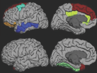
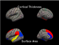
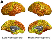
Using large scale studies, the authors assessed cortical measures in ADHD patients, unaffected siblings, and controls in adults, adolescents, and children. They found that the largest effects were in the surface areas of the frontal, cingulate, and temporal regions with some effect on thickness in the fusiform gyrus and temporal pole in children with no effects observed in adolescent or adult groups.
White Matter Microstructure and its Relation to Clinical Features of Obsessive-Compulsive Disorder: Findings from the ENIGMA OCD Working Group
Fabrizio Piras, Federica Piras, Yoshinari Abe, Sri Mahavir Agarwal, et al.
- Alan Anticevic, Stephanie Ameis, Paul Arnold, Núria Bargalló, Marcelo C. Batistuzzo, Francesco Benedetti, Jan-Carl Beucke, Premika S.W. Boedhoe, Irene Bollettini, Silvia Brem, Anna Calvo, Kang Ik Kevin Cho, Sara Dallaspezia, Erin Dickie, Benjamin Adam Ely, Siyan Fan, Jean-Paul Fouche, Patricia Gruner, Deniz A. Gürsel, Tobias Hauser, Yoshiyuki Hirano, Marcelo Q. Hoexter, Mariangela Iorio, Anthony James, Janardhan Reddy, Christian Kaufmann, Kathrin Koch, Peter Kochunov, Jun Soo Kwon, Luisa Lazaro, Christine Lochner, Rachel Marsh, Akiko Nakagawa, Takashi Nakamae, Janardhanan C. Narayanaswamy, Yuki Sakai, Eiji Shimizu, Daniela Simon, Helen Blair Simpson, Noam Soreni, Philipp Stämpfli, Emily R. Stern, Philip Szeszko, Jumpei Takahashi, Ganesan Venkatasubramanian, Zhen Wang, Je-Yeon Yun, ENIGMA OCD Working Group, Dan J. Stein, Neda Jahanshad, Paul M. Thompson, Odile A. van den Heuvel, Gianfranco Spalletta, and the ENIGMA OCD Working Group
The ENIGMA-OCD Working Group used DTI data from 1345 adults and 318 children from 19 sites to analyze white matter microstructural differences in OCD patients. They found significantly reduced FA values in the sagittal stratum and posterior thalamic radiation in adults. The lower FA was associated with longer duration of illness and early onset of OCD. There were no white matter changes found in pediatric patients.
Altered structural brain asymmetry in autism spectrum disorder in a study of 54 datasets
Merel C. Postema, Daan van Rooij, Evdokia Anagnostou, Celso Arango, et al.
- Guillaume Auzias, Marlene Behrmann, Geraldo Busatto Filho, Sara Calderoni, Rosa Calvo, Eileen Daly, Christine Deruelle, Adriana Di Martino, Ilan Dinstein, Fabio Luis S. Duran, Sarah Durston, Christine Ecker, Stefan Ehrlich, Damien Fair, Jennifer Fedor, Xin Feng, Jackie Fitzgerald, Dorothea L. Floris, Christine M. Freitag, Louise Gallagher, David C. Glahn, Ilaria Gori, Shlomi Haar, Liesbeth Hoekstra, Neda Jahanshad, Maria Jalbrzikowski, Joost Janssen, Joseph A. King, Xiang Zhen Kong, Luisa Lazaro, Jason P. Lerch, Beatriz Luna, Mauricio M. Martinho, Jane McGrath, Sarah E. Medland, Filippo Muratorj, Clodagh M. Murphy, Declan G.M. Murphy, Kirsten O’Hearn, Bob Oranje, Mara Parellada, Olga Puig, Alessandra Retico, Pedro Rosa, Katya Rubia, Devon Shook, Margot J. Taylor, Michela Tosetti Gregory L. Wallace, Fengfeng Zhou, Paul M. Thompson, Simon E. Fisher, Jan K. Buitelaar Clyde Francks
This study found thickness asymmetry associated with ASD in the medial frontal, orbitofrontal, cingulate and inferior temporal areas as well as asymmetry in the surface area of the orbitofrontal cortex. This generally manifests as reduced symmetry in ASD patients compared to healthy controls. There was also significant increased asymmetry in the putamen of ASD patients. This indicates that altered lateralization may be a feature of ASD.

No Alterations of Brain Structural Asymmetry in Major Depressive Disorder: An ENIGMA Consortium Analysis
Carolien G.F. de Kovel, Lyubomir Aftanas, André Aleman, Aaron F. Alexander-Bloch, et al.
- Bernhard T. Baune, Ivan Brack, Robin Bülow, Geraldo Busatto Filho, Angela Carballedo, Colm G. Connolly, Kathryn R. Cullen, Udo Dannlowski, Christopher G. Davey, Danai Dima, Katharina Dohm, Tracy Erwin-Grabner, Thomas Frodl, Cynthia H.Y. Fu, Geoffrey B. Hall, David C. Glahn, Beata Godlewska, Ian H. Gotlib, Roberto Goya-Maldonado, Hans Jörgen Grabe, Nynke A. Groenewold, Dominik Grotegerd, Oliver Gruber, Mathew A. Harris, Ben J. Harrison, Sean N. Hatton, Ian B. Hickie, Tiffany C. Ho, Neda Jahanshad, Tilo Kircher, Bernd Krämer, Axel Krug, Jim Lagopoulos, Elisabeth J. Leehr, Meng Li, Frank P. MacMaster, Glenda MacQueen, Andrew M. McIntosh, Quinn McLellan, Sarah E. Medland, Bryon A. Mueller, Igor Nenadic, Evgeny Osipov, Martina Papmeyer, Maria J. Portella, Liesbeth Reneman, Pedro G.P. Rosa, Matthew D. Sacchet, Knut Schnell, Anouk Schrantee, Kang Sim, Egle Simulionyte, Lisa Sindermann, Aditya Singh, M.S Dan J. Stein, Benjamin N. Ubani, Nic J.A. Van der Wee, Steven J.A. Van der Werff, Ilya M. Veer, Yolanda Vives-Gilabert, Henry Völzke, Henrik Walter, Martin Walter, Melinda Westlund Schreiner, Heather Whalley, Nils Winter, Katharina Wittfeld, Tony T. Yang, Dilara Yüksel, Dario Zaremba, Paul M. Thompson, Dick J. Veltman, Lianne Schmaal, Clyde Francks
This study investigated asymmetries of the brain using MRI data from about 2250 patients with Major Depressive Disorder and more than 3500 healthy controls. The largest effects asymmetries were observed in the thickness of the superior temporal cortex but was not significant when adjusting for multiple testing. There were no major differences in asymmetry between patients and controls.
Mapping Cortical and Subcortical Asymmetry in Obsessive-Compulsive Disorder: Findings From the ENIGMA Consortium
Xiang-Zhen Kong, Premika S.W. Boedhoe, Yoshinari Abe, Pino Alonso, et al.
- Stephanie H. Ameis, Paul D. Arnold, Francesca Assogna, Justin T. Baker, Marcelo C. Batistuzzo, Francesco Benedetti, Jan C. Beucke, Irene Bollettini, Anushree Bose, Silvia Brem, Brian P. Brennan, Jan Buitelaar, Rosa Calvo, Yuqi Cheng, Kang Ik K. Cho, Sara Dallaspezia, Damiaan Denys, Benjamin A. Ely, Jamie Feusner, Kate D. Fitzgerald, Jean-Paul Fouche, Egill A. Fridgeirsson, David C. Glahn, Patricia Gruner, Deniz A. Gürsel, Tobias U. Hauser, Yoshiyuki Hirano, Marcelo Q. Hoexter, Hao Hu, Chaim Huyser, Anthony James, Fern Jaspers-Fayer, Norbert Kathmann, Christian Kaufmann, Kathrin Koch, Masaru Kuno, Gerd Kvale, Jun Soo Kwon, Luisa Lazaro, Yanni Liu, Christine Lochner, Paulo Marques, Rachel Marsh, Ignacio Martínez-Zalacaín, David Mataix-Cols, Sarah E. Medland,José M. Menchón, Luciano Minuzzi, Pedro S. Moreira, Astrid Morer, Pedro Morgado,Akiko Nakagawa, Takashi Nakamae, Tomohiro Nakao, Janardhanan C. Narayanaswamy, Erika L. Nurmi, Joseph O’Neill, Jose C. Pariente, Chris Perriello, John Piacentini, Fabrizio Piras, Federica Piras, Christopher Pittenger, Y.C. Janardhan Reddy, Oana Georgiana Rus-Oswald, Yuki Sakai, Joao R. Sato, Lianne Schmaal, H. Blair Simpson, Noam Soreni, Carles Soriano-Mas, Gianfranco Spalletta, Emily R. Stern, Michael C. Stevens, S. Evelyn Stewart, Philip R. Szeszko, David F. Tolin, Aki Tsuchiyagaito, Daan van Rooij, Guido A. van Wingen, Ganesan Venkatasubramanian, Zhen Wang, Je-Yeon Yun, ENIGMA OCD Working Group, Paul M. Thompson, Dan J. Stein, Odile A. van den Heuvel, and Clyde Francks
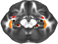
The OCD Working Group of the ENIGMA Consortium analyzed the cortical thickness, cortical surface area, and subcortical volumes of 940 child and 3431 adult participants focusing on brain asymmetry. They found the largest differences between asymmetry in OCD patients and controls were in volumes of the thalamus and pallidum in children, which suggest subtle changes in the asymmetry of subcortical regions in children not visible in adults.
Mapping cortical brain asymmetry in 17,141 healthy individuals worldwide via the ENIGMA Consortium
Xiang-Zhen Kong, Samuel R. Mathias, Tulio Guadalupe, et al.
- ENIGMA Laterality Working Group, David C. Glahn, Barbara Franke, Fabrice Crivello, Nathalie Tzourio-Mazoyer, Simon E. Fisher, Paul M. Thompson, and Clyde Francks
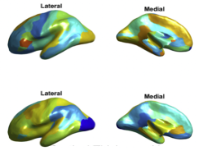
With 17,141 participants ENIGMA Consortium presents the largest study of cortical asymmetry focusing on cortical thickness and surface area variability. They found wide-spread asymmetries in thicker cortices but smaller surface areas in the left hemisphere. There were also regional asymmetries mainly in regions involved in lateralized functions. Variability in brain asymmetry was also found to be related to sex, age, and intracranial volumes, and was found to be significantly heritable.
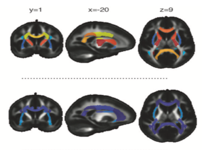
White matter disturbances in major depressive disorder:a coordinated analysis across 20 international cohorts inthe ENIGMA MDD working group
Laura S. van Velzen, Sinead Kelly, Dmitry Isaev, Andre Aleman, et al.
- Lyubomir I. Aftanas, Jochen Bauer, Bernhard T. Baune, Ivan V. Brak, Angela Carballedo, Colm G. Connolly, Baptiste Couvy-Duchesne, Kathryn R. Cullen, Konstantin V. Danilenko, Udo Dannlowski, Verena Enneking, Elena Filimonova, Katharina Förster, Thomas Frodl, Ian H. Gotlib, Nynke A. Groenewold, Dominik Grotegerd, Mathew A. Harris, Sean N. Hatton, Emma L. Hawkins, Ian B. Hickie, Tiffany C. Ho, Andreas Jansen, Tilo Kircher, Bonnie Klimes-Dougan, Peter Kochunov, Axel Krug, Jim Lagopoulos, Renick Lee, Tristram A. Lett, Meng Li, Frank P. MacMaster, Nicholas G. Martin, Andrew M. McIntosh, Quinn McLellan, Susanne Meinert, Igor Nenadić, Evgeny Osipov, Brenda W. J. H. Penninx, Maria J. Portella, Jonathan Repple, Annerine Roos, Matthew D. Sacchet, Philipp G. Sämann, Knut Schnell, Xueyi Shen, Kang Sim, Dan J. Stein, Marie-Jose van Tol, Alexander S. Tomyshev, Leonardo Tozzi, Ilya M. Veer, Robert Vermeiren, Yolanda Vives-Gilabert, Henrik Walter, Martin Walter, Nic J. A. van der Wee, Steven J. A. van der Werff, Melinda Westlund Schreiner, Heather C. Whalley, Margaret J. Wright, Tony T. Yang, Alyssa Zhu, Dick J. Veltman, Paul M. Thompson, Neda Jahanshad, Lianne Schmaal
White matter anisotropy and diffusivity data were collected from 1305 Major Depressive Disorder (MDD) patients and 1602 healthy controls from 20 samples from around the world in the ENIGMA consortium. Effect sizes were meta-analyzed across studies and showed widespread lower fractional anisotropy in adult patients when compared to controls in 16 of 25 regions of interest. The largest differences were seen in the corpus callosum and corona radiata. White matter microstructural differences between adolescent patients and controls were not apparent after correction for multiple testing. These findings may suggest structural disconnectivity in MDD.
Widespread White Matter Microstructural Abnormalities in Bipolar Disorder: Evidence from mega- and meta-analyses across 3,033 individuals
Pauline Favre, Melissa Pauling, Jacques Stout, Franz Hozer, et al.
- Samuel Sarrazin, Christoph Abé, Martin Alda, Clara Alloza, Silvia Alonso-Lana, Ole A Andreassen, Bernhard T Baune, Francesco Benedetti, Geraldo F Busatto, Erick J Canales-Rodríguez, Xavier Caseras, Tiffany Moukbel Chaim-Avancini, Christopher R.K. Ching, Udo Dannlowski, Michael Deppe, Lisa T Eyler, Mar Fatjo-Vilas, Sonya F Foley, Dominik Grotegerd, Tomas Hajek, Unn K Haukvik, Fleur M Howells, Neda Jahanshad, Harald Kugel, Trine V Lagerberg, Stephen M Lawrie, Julia O Linke, Andrew McIntosh, Elisa M.T. Melloni, Philip B Mitchell, Mircea Polosan, Edith Pomarol-Clotet, Jonathan Repple, Gloria Roberts, Annerine Roos, Pedro G.P. Rosa, Raymond Salvador, Salvador Sarró, Peter R Schofield, Mauricio H Serpa, Kang Sim, Dan J Stein, Jess E Sussmann, Henk S Temmingh, Paul M Thompson, Norma Verdolini, Eduard Vieta, Michele Wessa, Heather C Whalley, Marcus V Zanetti, Marion Leboyer, Jean-François Mangin, Chantal Henry, Edouard Duchesnay, Josselin Houenou, for the ENIGMA Bipolar Disorder Working Group
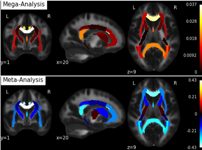
Using the largest bipolar disorder diffusion tensor imaging dataset to date, mega- and meta-analyses were conducted comparing images from bipolar disorder patients with those from healthy controls. The data were gathered from 26 cohorts, resulting in a sample size of N = 3,033. The analyses showed significantly lowered FA in patients in the corpus callosum and cingulum with lithium medication, later onset and short disease duration also related to higher FA in multiple regions.
Altered White Matter Microstructural Organization in Post-Traumatic Stress Disorder across 3,049 Adults: Results from the PGC-ENIGMA PTSD Consortium
Emily L Dennis, Seth G Disner, Negar Fani, Lauren E Salminen, et al.
- Mark Logue, Emily K Clarke, Courtney C Haswell, Christopher L Averill, Lee A Baugh, Jessica Bomyea, Steven E Bruce, Jiook Cha, Kyle Choi, Nicholas D Davenport, Maria Densmore, Stefan du Plessis, Gina L Forster, Jessie L Frijling, Atilla Gönenc, Staci Gruber, Daniel W Grupe, Jeffrey P Guenette, Jasmeet Hayes, David Hofmann, Jonathan Ipser, Tanja Jovanovic, Sinead Kelly, Mitzy Kennis, Philipp Kinzel, Saskia BJ Koch, Inga Koerte, Sheri Koopowitz, Mayuresh Korgaonkar, John Krystal, Lauren AM Lebois, Gen Li, Vincent A Magnotta, Antje Manthey, GeoffreyJ May, Deleene S Menefee, Laura Nawijn, Steven M Nelson, Richard WJ Neufeld, Jack B Nitschke, Daniel O'Doherty, Matthew Peverill, Kerry Ressler, Annerine Roos, Margaret A Sheridan, Anika Sierk, Alan Simmons, Raluca M Simons, Jeffrey S Simons, Jennifer Stevens, Benjamin Suarez-Jimenez, Danielle R Sullivan, Jean Théberge, Jana K Tran, Leigh van den Heuvel, Steven JA van der Werff, Sanne JH van Rooij, Mirjam van Zuiden, Carmen Velez, Mieke Verfaellie, Robert RJM Vermeiren, Benjamin SC Wade, Tor Wager, Henrik Walter, Sherry Winternitz, Jonathan Wolff, Gerald York, Ye Zhu, Xi Zhu, Chadi G Abdallah, Richard Bryant, Judith K Daniels, Richard J Davidson, Kelene A Fercho, Carol Franz, Elbert Geuze, Evan M Gordon, Milissa L Kaufman, William Kremen, Jim Lagopoulos, Ruth A Lanius, Michael J Lyons, Stephen R McCauley, Regina McGlinchey, Katie A McLaughlin, William Milberg, Yuval Neria, Miranda Olff, Soraya Seedat, Martha Shenton, Scott R Sponheim, Dan J Stein, Murray B Stein, Thomas Straube, David F Tate, Nic JA van der Wee, Dick J Veltman, Li Wang, Elisabeth A Wilde, Paul M Thompson, Peter Kochunov, Neda Jahanshad, Rajendra A Morey
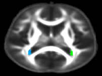
In this study the diffusion MRI data of 1446 individuals with PTSD and 1603 controls from 28 cohorts in the PGC-ENIGMA PTSF working group were analyzed for differences in white matter tracts. The analysis results showed lower fractional anisotropy in the tapetum region of the corpus callosum, which connects the left and right hippocampus, structures previously implicated in PTSD. These results support an association between PTSD and alterations of the broader hippocampal network.
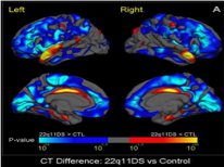
Altered White Matter Microstructure in 22q11.2 Deletion Syndrome: A Multisite Diffusion Tensor Imaging Study
Julio E. Villalón-Reina, Kenia Martínez, Xiaoping Qu, Christopher R. K. Ching, et al.
- Talia M. Nir, Deydeep Kothapalli, Conor Corbin, Daqiang Sun, Amy Lin, Jennifer K. Forsyth, Leila Kushan, Ariana Vajdi, Maria Jalbrzikowski, Laura Hansen, Rachel K. Jonas, Therese van Amelsvoort, Geor Bakker, Wendy R. Kates, Kevin M. Antshel, Wanda Fremont, Linda E. Campbell, Kathryn L. McCabe, Eileen Daly, Maria Gudbrandsen, Clodagh M. Murphy, Declan Murphy, Michael Craig, Beverly Emanuel, Donna M. McDonald-McGinn, Jacob A.S. Vorstman, Ania M. Fiksinski, Sanne Koops, Kosha Ruparel, David Roalf, Raquel E. Gur, J. Eric Schmitt, Tony J. Simon, Naomi J. Goodrich-Hunsaker, Courtney A. Durdle, Joanne L. Doherty, Adam C. Cunningham, Marianne van den Bree, David E. J. Linden, Michael Owen, Hayley Moss, Sinead Kelly, Gary Donohoe, Kieran C. Murphy, Celso Arango, Neda Jahanshad, Paul M. Thompson, Carrie E. Bearden
Meta- and mega- analyses were performed on DTI scans of 334 22q11.2 deletion carriers and 260 healthy controls in the largest dMRI0-dervied measures of white matter microstructure in 22q11.2 Deletion Syndrome yet. The analyses results indicated major reductions in mean, axial, and radial diffusivities in 22q11DS patients in the corona radiata, corpus callosum, superior longitudinal fasciculus, posterior thalamic radiations and sagittal stratum. However, 22q11DS patients showed higher mean fractional anisotropy (FA) in the internal capsule and corona radiata and lower FA in association fibers compared to controls.
Cortical Abnormalities Associated with Pediatric and Adult Obsessive-Compulsive Disorder: Findings from the ENIGMA Obsessive-Compulsive Disorder Working Group
Premika S.W. Boedhoe, Lianne Schmaal, Yoshinari Abe, Pino Alonso, et al.
- Stephanie H. Ameis, Alan Anticevic, Paul D. Arnold, Marcelo C. Batistuzzo, Francesco Benedetti, Jan C. Beucke, Irene Bollettini, Anushree Bose, Silvia Brem, Anna Calvo, Rosa Calvo, Yuqi Cheng, Kang Ik K. Cho, Valentina Ciullo, Sara Dallaspezia, Damiaan Denys, Jamie D. Feusner, Kate D. Fitzgerald, Jean-Paul Fouche, Egill A. Fridgeirsson, Patricia Gruner, Gregory L. Hanna, Derrek P. Hibar, Marcelo Q. Hoexter, Hao Hu, Chaim Huyser, Neda Jahanshad, Anthony James, Norbert Kathmann, Christian Kaufmann, Kathrin Koch, Jun Soo Kwon, Luisa Lazaro, Christine Lochner, Rachel Marsh, Ignacio Martínez-Zalacaín, David Mataix-Cols, José M. Menchón, Luciano Minuzzi, Astrid Morer, Takashi Nakamae, Tomohiro Nakao, Janardhanan C. Narayanaswamy, Seiji Nishida, Erika Nurmi, Joseph O’Neill, John Piacentini, Fabrizio Piras, Federica Piras, Y.C. Janardhan Reddy, Tim J. Reess, M.A., Yuki Sakai, Joao R. Sato, H. Blair Simpson, Noam Soreni, Carles Soriano-Mas, Gianfranco Spalletta, Michael C. Stevens, Philip R. Szeszko, David F. Tolin, Guido A. van Wingen, Ganesan Venkatasubramanian, Susanne Walitza, Zhen Wang, Je-Yeon Yun, ENIGMA-OCD Working Group, Paul M. Thompson, Dan J. Stein, Odile A. van den Heuvel
Using T1-weighted MRI scans of 1,905 patients with obsessive-compulsive disorder (OCD) and 1,760 healthy controls from 27 centers worldwide, the authors conducted meta- and mega-analyses to study structural abnormalities of the brain in OCD patients. They found that the inferior parietal cortex was significantly thinner in patients when compared to controls, and that the inferior and superior parietal cortices were thinner in medicated pediatric patients than in non-medicated pediatric patients compared to controls, indicating that these cortical measures may be affected by the different stages of illness and development.
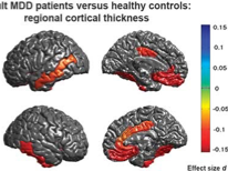
Cortical abnormalities in adults and adolescents with major depression based on brain scans from 20 cohorts worldwide in the ENIGMA Major Depressive Disorder Working Group
L Schmaal, DP Hibar, PG Sämann, GB Hall, et al.
- BT Baune, N Jahanshad, JW Cheung, TGM van Erp, D Bos, MA Ikram, MW Vernooij, WJ Niessen, H Tiemeier, A Hofman, K Wittfeld, HJ Grab, D Janowitz, R Bülow, M Selonke, H Völzke, D Grotegerd, U Dannlowski, V Arolt, N Opel, W Heindel, H Kugel, D Hoehn, M Czisch, B Couvy-Duchesne, ME Rentería, LT Strike, MJ Wright, NT Mills, GI de Zubicaray, KL McMahon, SE Medland, NG Martin, NA Gillespie, R Goya-Maldonado, O Gruber, B Krämer, SN Hatton, J Lagopoulos, IB Hickie, T Frodl, A Carballedo, EM Frey, LS van Velzen, BWJH Penninx, M-J van Tol, NJ van der Wee, CG Davey, BJ Harrison, B Mwangi, B Cao, JC Soares, IM Veer, H Walter, D Schoepf, B Zurowski, C Konrad, E Schramm, C Normann, K Schnell Sacchet, IH Gotlib, GM MacQueen, BR Godlewska, T Nickson, AM McIntosh, M Papmeyer, HC Whalley, J Hall, JE Sussmann, M Li, M Walter, L Aftanas, I Brack, NA Bokhan, PM Thompsom and DJ Veltman for the ENIGMA-Major Depressive Disorder Working Group
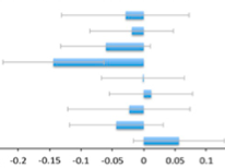
The ENIGMA Major Depressive Disorder (MDD) Working Group conducted meta-analyses on the T1-weighted MRI scans of 2,148 MDD patients and 7,957 healthy controls from 20 sites around the world, analyzing adults and adolescents separately. Adults patients were found to have thinner grey matter in the orbitofrontal cortex, anterior and posterior cingulate, insula, and temporal lobes compared to healthy controls. Adolescent patients had lower surface areas, with effects greatest among recurrent patients. These findings indicate consistent cortical alterations in patients compared to controls and imply that MDD may affect brain structure in different ways depending on age.
Large-scale mapping of cortical alterations in 22q11.2 deletion syndrome: Convergence with idiopathic psychosis and effects of deletion size
Daqiang Sun, Christopher R. K. Ching, Amy Lin, Jennifer K. Forsyth, et al.
- Leila Kushan, Ariana Vajdi, Maria Jalbrzikowski, Laura Hansen, Julio E. Villalon-Reina, Xiaoping Qu, Rachel K. Jonas, Therese van Amelsvoort, Geor Bakker, Wendy R. Kates, Kevin M. Antshel, Wanda Fremont, Linda E. Campbell, Kathryn L. McCabe, Eileen Daly, Maria Gudbrandsen, Clodagh M. Murphy, Declan Murphy, Michael Craig, Jacob Vorstman, Ania Fiksinski, Sanne Koops, Kosha Ruparel, David R. Roalf, Raquel E. Gur, J. Eric Schmitt, Tony J. Simon, Naomi J. Goodrich-Hunsaker, Courtney A. Durdle, Anne S. Bassett, Eva W. C. Chow, Nancy J. Butcher, Fidel Vila-Rodriguez, Joanne Doherty, Adam Cunningham, Marianne B.M. van den Bree, David E. J. Linden, Hayley Moss, Michael J. Owen, Kieran C. Murphy, Donna M. McDonald-McGinn, Beverly Emanuel, Theo G. M. van Erp, Jessica A. Turner, Paul M. Thompson, Carrie E. Bearden
This study consisted of meta-analyses of data from 474 individuals with the 22q11.2 chromosomal microdeletion (22q11DS) risk factor for psychotic illness and 315 individuals developing typically gathered from 10 sites around the world. Overall, grey matter thickness was greater in the 22q11DS subjects, but there were focal thickness reductions in the temporal and cingulate cortices. There were global reductions of cortical surface area in 22q11DS individuals compared to controls, and there were significant differences in the reduction based on the size of the 22q11DS deletion (1.5 Mb or 3 Mb). The 22q11DS subjects showed brain structure similarities when compared with idiopathic schizophrenia, particularly in the fronto-temporal cortex, which supports the suitability of 22q11DS as a biological model of schizophrenia.
Cortical Brain Abnormalities in 4474 Individuals with Schizophrenia and 5098 Control Subjects via the Enhancing Neuro Imaging Genetics Through Meta-Analysis (ENIGMA) Consortium
Theo G.M. van Erp, Esther Walton, Derrek P. Hibar, Lianne Schmaal, et al.
- Wenhao Jiang, David C. Glahn, Godfrey D. Pearlson, Nailin Yao, Masaki Fukunaga, Ryota Hashimoto, Naohiro Okada, Hidenaga Yamamori, Juan R. Bustillo, Vincent P. Clark, Ingrid AgartzIngrid Agartz, Bryon A. Mueller, Wiepke Cahn, Sonja M.C. de Zwarte, Hilleke E. Hulshoff Pol, René S. Kahn, Roel A. Ophoff, Neeltje E.M. van Haren, Ole A. Andreassen, Anders M. Dale, Nhat Trung Doan, Tiril P. Gurholt, Cecilie B. Hartberg, Unn K. Haukvik, Kjetil N. Jørgensen, Trine V. Lagerberg, Ingrid Melle, Lars T. Westlye, Oliver Gruber, Bernd Kraemer, Anja Richter, David Zilles, Vince D. Calhoun, Benedicto Crespo-Facorro, Roberto Roiz-Santiañez, Diana Tordesillas-Gutiérrez, Carmel Loughland, Vaughan J. Carr, Stanley Catts, Vanessa L. Cropley, Janice M. Fullerton, Melissa J. Green, Frans A. Henskens, Assen Jablensky, Rhoshel K. Lenroot, Bryan J. Mowry, Patricia T. Michie, Christos Pantelis, Yann Quidé, Ulrich Schall, Rodney J. Scott, Murray J. Cairns, Marc Seal, Paul A. Tooney, Paul E. Rasser, Gavin Cooper, Cynthia Shannon Weickert, Thomas W. Weickert, Derek W. Morris, Elliot Hong, Peter Kochunov, Lauren M. Beard, Raquel E. Gur, Ruben C. Gur, Theodore D. Satterthwaite, Daniel H. Wolf, Aysenil Belger, Gregory G. Brown, Judith M. Ford, Fabio Macciardi, Daniel H. Mathalon, Daniel S. O’Leary, Steven G. Potkin, Adrian Preda, James Voyvodic, Kelvin O. Lim, Sarah McEwen, Fude Yang, Yunlong Tan, Shuping Tan, Zhiren Wang, Fengmei Fan, Jingxu Chen, Hong Xiang, Shiyou Tang, Hua Guo, Ping Wan, Dong Wei, Henry J. Bockholt, Stefan Ehrlich, Rick P.F. Wolthusen, Margaret D. King, Jody M. Shoemaker, Scott R. Sponheim, Lieuwe De Haan, Laura Koenders, Marise W. Machielsen, Therese van Amelsvoort, Dick J. Veltman, Francesca Assogna, Nerisa Banaj, Pietro de Rossi, Mariangela Iorio, Fabrizio Piras, Gianfranco Spalletta, Peter J. McKenna, Edith Pomarol-Clotet, Raymond Salvador, Aiden Corvin, Gary Donohoe, Sinead Kelly, Christopher D. Whelan, Erin W. Dickie, David Rotenberg, Aristotle N. Voineskos, Simone Ciufolini, Joaquim Radua, Paola Dazzan, Robin Murray, Tiago Reis Marques, Andrew Simmons, Stefan Borgwardt, Laura Egloff, Fabienne Harrisberger, Anita Riecher-Rössler, Renata Smieskova, Kathryn I. Alpert, Lei Wang, Erik G. Jönsson, Sanne Koops, Iris E.C. Sommer, Alessandro Bertolino, Aurora Bonvino, Annabella Di Giorgio, Emma Neilson, Andrew R. Mayer, Julia M. Stephen, Jun Soo Kwon, Je-Yeon Yun, Dara M. Cannon, Colm McDonald, Irina Lebedeva, Alexander S. Tomyshev, Tolibjohn Akhadov, Vasily Kaleda, Helena Fatouros-Bergman, Lena FlycktLena FlycktHelena Fatouros-Bergman, Lena Flyckt, Karolinska Schizophrenia Project, Geraldo F. Busatto, Pedro G.P. Rosa, Mauricio H. Serpa, Marcus V. Zanetti, Cyril Hoschl, Antonin Skoch, Filip Spaniel, David Tomecek, Saskia P. Hagenaars, Andrew M. McIntosh, Heather C. Whalley, Stephen M. Lawrie, Christian Knöchel, Viola Oertel-Knöchel, Michael Stäblein, Fleur M. Howells, Dan J. Stein, Henk S. Temmingh, Anne Uhlmann, Carlos Lopez-Jaramillo, Danai Dima, Agnes McMahon, Joshua I. Faskowitz, Boris A. Gutman, Neda Jahanshad, Paul M. Thompson, and Jessica A. Turner
This study used data from 4,474 patients with schizophrenia and 5,098 healthy controls from 39 centers around the world to conduct a meta-analysis of cortical thickness and surface area abnormalities in patients. Patients with schizophrenia had smaller surface areas especially in the frontal and temporal lobes and had regionally specific thinner cortices. The cortical thickness effect sizes greatly depended on medication, duration of illness, age of onset, indicating that genetic association studies of schizophrenia and cortical thickness should take these variables into account.
Subcortical brain volume differences in participants with attention deficit hyperactivity disorder in children and adults: a cross-sectional mega-analysis
Martine Hoogman, Janita Bralten, Derrek P Hibar, Maarten Mennes, et al.
- Marcel P Zwiers, Lizanne S J Schweren, Kimm J E van Hulzen, Sarah E Medland, Elena Shumskaya, Neda Jahanshad, Patrick de Zeeuw, Eszter Szekely, Gustavo Sudre, Thomas Wolfers, Alberdingk M H Onnink, Janneke T Dammers, Jeanette C Mostert, Yolanda Vives-Gilabert, Gregor Kohls, Eileen Oberwelland, Jochen Seitz, Martin Schulte-Rüther, Sara Ambrosino, Alysa E Doyle, Marie F Høvik, Margaretha Dramsdahl, Leanne Tamm, Theo G M van Erp, Anders Dale, Andrew Schork, Annette Conzelmann, Kathrin Zierhut, Ramona Baur, Hazel McCarthy, Yuliya N Yoncheva, Ana Cubillo, Kaylita Chantiluke, Mitul A Mehta, Yannis Paloyelis, Sarah Hohmann, Sarah Baumeister, Ivanei Bramati, Paulo Mattos, Fernanda Tovar-Moll, Pamela Douglas, Tobias Banaschewski, Daniel Brandeis, Jonna Kuntsi, Philip Asherson, Katya Rubia, Clare Kelly, Adriana Di Martino, Michael P Milham, Francisco X Castellanos, Thomas Frodl, Mariam Zentis, Klaus-Peter Lesch, Andreas Reif, Paul Pauli, Terry L Jernigan, Jan Haavik, Kerstin J Plessen, Astri J Lundervold, Kenneth Hugdahl, Larry J Seidman, Joseph Biederman, Nanda Rommelse, Dirk J Heslenfeld, Catharina A Hartman, Pieter J Hoekstra, Jaap Oosterlaan, Georg von Polier, Kerstin Konrad, Oscar Vilarroya, Josep Antoni Ramos-Quiroga, Joan Carles Soliva, Sarah Durston, Jan K Buitelaar, Stephen V Faraone, Philip Shaw, Paul M Thompson, Barbara Franke
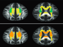
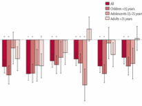
The ENIGMA attention deficit hyperactivity disorder (ADHD) Working Group conducted mega-analyses comparing the T1-weighted MRI scans of 1,713 participants with ADHD with data from 1,529 control participants. They found that the volumes of the accumbens, amygdala, caudate, hippocampus, and putamen in participants with ADHD were significantly smaller than in controls. The analysis also showed a greatest effect sizes in participants under the age of 15 compared to adults.
Distinct Subcortical Volume Alterations in Pediatric and Adult OCD: A Worldwide Meta- and Mega-Analysis
Premika S.W. Boedhoe, Lianne Schmaal, Yoshinari Abe, Stephanie H. Ameis, et al.
- Paul D. Arnold, Marcelo C. Batistuzzo, Francesco Benedetti, Jan C. Beucke, Irene Bollettini, Psy.D., Anushree Bose, M.A., Silvia Brem, Anna Calvo, Yuqi Cheng, Kang Ik K. Cho, B.Sc., Sara Dallaspezia, Damiaan Denys, Kate D. Fitzgerald, Jean-Paul Fouche, Mònica Giménez, Patricia Gruner, Gregory L. Hanna, Derrek P. Hibar, Marcelo Q. Hoexter, Hao Hu, Chaim Huyser, Keisuke Ikari, Neda Jahanshad, Norbert Kathmann, Christian Kaufmann, Kathrin Koch, Jun Soo Kwon, Luisa Lazaro, Yanni Liu, Christine Lochner, Rachel Marsh, Ignacio Martínez-Zalacaín, David Mataix Cols, José M. Menchón, Luciano Minuzzi, Takashi Nakamae, Tomohiro Nakao, Janardhanan C. Narayanaswamy, Fabrizio Piras, Federica Piras, Christopher Pittenger, Y.C. Janardhan Reddy, Joao R. Sato, H. Blair Simpson, Noam Soreni, Carles Soriano-Mas, Gianfranco Spalletta, Michael C. Stevens, Philip R. Szeszko, David F. Tolin, Ganesan Venkatasubramanian, Susanne Walitza, Zhen Wang, Guido A. van Wingen, Jian Xu, Xiufeng Xu, Je-Yeon Yun, Qing Zhao, ENIGMA OCD Working Group, Paul M. Thompson, Dan J. Stein, Odile A. van den Heuvel
Mega- and meta- analyses were conducted on the T1-weighted images from 1,830 patients with obsessive-compulsive disorder (OCD) and 1,759 health controls. The results indicated that patients had significantly smaller hippocampal volumes and larger pallidum volumes compared to controls, and that the effects were stronger in medicated patients. Pediatric patients also had significantly larger thalamic volumes. These results indicate a difference in abnormalities of subcortical volumes between adult and pediatric OCD patients.
Subcortical brain volume abnormalities in 2028 individuals with schizophrenia and 2540 healthy controls via the ENIGMA consortium
TGM van Erp, DP Hibar, JM Rasmussen, DC Glahn, et al.
- GD Pearlson, OA Andreassen, I Agartz, LT Westlye, UK Haukvik, AM Dale, I Melle, CB Hartberg, O Gruber, B Kraemer, D Zilles, G Donohoe, S Kelly, C McDonald, DW Morris, DM Cannon, A Corvin, MWJ Machielsen, L Koenders, L de Haan, DJ Veltman, TD Satterthwaite, DH Wolf, RC Gur, RE Gur, SG Potkin, DH Mathalon, BA Mueller, A Preda, F Macciardi, S Ehrlich, E Walton, Hass, VD Calhoun, HJ Bockholt, SR Sponheim, JM Shoemaker, NEM van Haren, HEH Pol, RA Ophoff, RS Kahn, R Roiz-Santiañez, B Crespo-Facorro, L Wang, KI Alpert, EG Jönsson, R Dimitrova, C Bois, HC Whalley, AM McIntosh, SM Lawrie, R Hashimoto, PM Thompson, and JA Turner, for the ENIGMA Schizophrenia Working Group
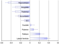
Using MRI scans from 2,028 schizophrenia patients and 2,540 healthy controls from 15 sites worldwide, researchers conducted neuroimaging data meta-analyses. They found that schizophrenia patients showed smaller hippocampal, amygdala, thalamic, accumbens, and intracranial volumes, but larger pallidum and lateral ventricle volumes.
Subcortical brain alterations in major depressive disorder: findings from the ENIGMA Major Depressive Disorder working group
L Schmaal, DJ Veltman, TGM van Erp, PG Sämann, et al.
- T Frodl, N Jahanshad, E Loehrer, H Tiemeier, A Hofman, WJ Niessen, MW Vernooij, MA Ikram, K Wittfeld, HJ Grabe, A Block, K Hegenscheid, H Völzke, D Hoehn, M Czisch, J Lagopoulos, SN Hatton, IB Hickie, R Goya-Maldonado, B Krämer, O Gruber, B Couvy-Duchesne, ME Rentería, LT Strike, NT Mills, GI de Zubicaray, KL McMahon, SE Medland, NG Martin, NA Gillespie, MJ Wright, GB Hall, GM MacQueen, EM Frey, A Carballedo, LS van Velzen, MJ van Tol, NJ van der Wee, IM Veer, H Walter, K Schnell, E Schramm, C Normann, D Schoepf, C Konrad, B Zurowski, T Nickson, AM McIntosh, M Papmeyer, HC Whalley, JE Sussmann, BR Godlewska, PJ Cowen, FH Fischer, M Rose, BWJH Penninx, PM Thompson, and DP Hibar for the ENIGMA-Major Depressive Disorder Working Group
This study consists of meta-analyses of MRI brain scans from 1,728 patients with major depressive disorder (MDD) and 7,199 healthy controls from 15 researcher centers around the world. The results indicate that relative to controls, patients have a smaller volume hippocampus, while later onset MDD was also associated with smaller amygdala and larger lateral ventricles.
Evidence for Widespread White Matter Microstructural Differences in Schizophrenia Across 4,375 Individuals from 30 International Studies: Results from the ENIGMA Schizophrenia DTI Working Group
We harmonized the processing of diffusion tensor imaging (DTI) data of 2391 healthy controls and 1,984 schizophrenia patients from 30 studies worldwide and meta-analyzed regional effects. We have observed significantly lower FA values for global and regional FA values. Longer duration of illness was significantly and negativly correlated with FA in subregions of the corpus collosum, external capsule, fornix, posterior thalamic radiation, superior longitudinal fasciculus and sagittal stratum.

Heterochronicity of White Matter Development and Aging and Vulnerability to Schizophrenia
Kochunov Peter, Ganjgahi Halib, Winkler Anderson, et al.
- Kochunov Peter, Ganjgahi Halib, Winkler Anderson, Kelly Sinead, Shukla Dinesh, Xiaoming Du, Jahanshad Neda, Rowland Laura, Sampath Hemalatha, Patel Binish, O’Donnell Patricio, Xie Zhiyong, Paciga Sara A, Schubert Christian, Chen Jian, Zhang Guohao, Thompson Paul M., Nichols Thomas E., Hong Elliot L.
Alternated brain connectivity has been implicated in the development and clinical burden of schizophrenia. We tested if differences in the trajectories of white matter tract development influenced the patient-control differences observed in FA and if in turn, those tracts show exacerbated decline with aging.
Structural brain abnormalities in the common epilepsies assessed in a worldwide ENIGMA study
Whelan CD, Altmann A, Botía JA, Jahanshad N, Hibar DP, et al.
- Christopher D Whelan, Andre Altmann, Juan A Botía, Neda Jahanshad, Derrek P Hibar, Julie Absil, Saud Alhusaini, Marina K M Alvim, Pia Auvinen, Emanuele Bartolini, Felipe P G Bergo, Tauana Bernardes, Karen Blackmon, Barbara Braga, Maria Eugenia Caligiuri, Anna Calvo, Sarah J Carr, Jian Chen, Shuai Chen, Andrea Cherubini, Philippe David, Martin Domin, Sonya Foley, Wendy França, Gerrit Haaker, Dmitry Isaev, Simon S Keller, Raviteja Kotikalapudi, Magdalena A Kowalczyk, Ruben Kuzniecky, Soenke Langner, Matteo Lenge, Kelly M Leyden, Min Liu, Richard Q Loi, Pascal Martin, Mario Mascalchi, Marcia E Morita, Jose C Pariente, Raul Rodríguez-Cruces, Christian Rummel, Taavi Saavalainen, Mira K Semmelroch, Mariasavina Severino, Rhys H Thomas, Manuela Tondelli, Domenico Tortora, Anna Elisabetta Vaudano, Lucy Vivash, Felix von Podewils, Jan Wagner, Bernd Weber, Yi Yao, Clarissa L Yasuda, Guohao Zhang, Nuria Bargalló, Benjamin Bender, Neda Bernasconi, Andrea Bernasconi, Boris C Bernhardt, Ingmar Blümcke, Chad Carlson, Gianpiero L Cavalleri, Fernando Cendes, Luis Concha, Norman Delanty, Chantal Depondt, Orrin Devinsky, Colin P Doherty, Niels K Focke, Antonio Gambardella, Renzo Guerrini, Khalid Hamandi, Graeme D Jackson, Reetta Kälviäinen, Peter Kochunov, Patrick Kwan, Angelo Labate, Carrie R McDonald, Stefano Meletti, Terence J O'Brien, Sebastien Ourselin, Mark P Richardson, Pasquale Striano, Thomas Thesen, Roland Wiest, Junsong Zhang, Annamaria Vezzani, Mina Ryten, Paul M Thompson, Sanjay M Sisodiya
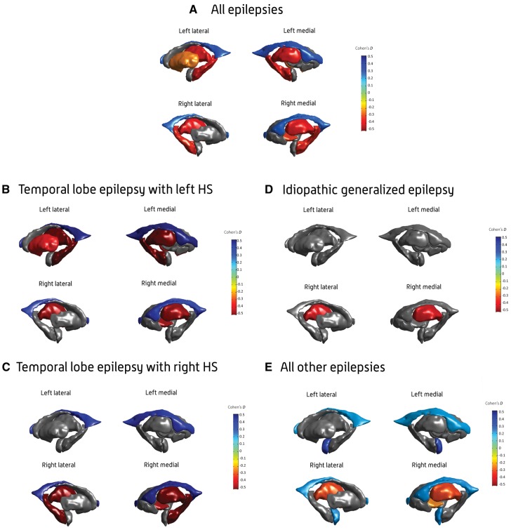
Progressive functional decline in the epilepsies is largely unexplained. We formed the ENIGMA-Epilepsy consortium to understand factors that influence brain measures in epilepsy, pooling data from 24 research centres in 14 countries across Europe, North and South America, Asia, and Australia. Structural brain measures were extracted from MRI brain scans across 2149 individuals with epilepsy, divided into four epilepsy subgroups including idiopathic generalized epilepsies (n =367), mesial temporal lobe epilepsies with hippocampal sclerosis (MTLE; left, n = 415; right, n = 339), and all other epilepsies in aggregate (n = 1026), and compared to 1727 matched healthy controls.
Alzheimer’s Disease-Like Brain Pattern Biomarker: Capturing Risks and Predicting Disease Onset
Peter Kochunov PhD., Si Gao MS, Lauren E. Salminen PhD., Neda Jahanshad PhD., et al.
- Peter Kochunov PhD., Si Gao MS, Lauren E. Salminen PhD., Neda Jahanshad PhD., Talia M. Nir PhD., Paul M. Thompson PhD., Xiaoming Du PhD., Bhim M. Adhikari PhD.,Alice Kochunov MD., Ryan Cassidy, MD, PhD., Yizhou Ma PhD. , Joshua Chiappelli MD., Seth Ament PhD., Yezhi Pan, Shuo Chen PhD, Alan R. Shuldiner MD, Braxton D. Mitchell PhD, L. Jair Soares MD., Elliot Hong MD.
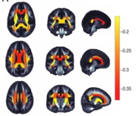
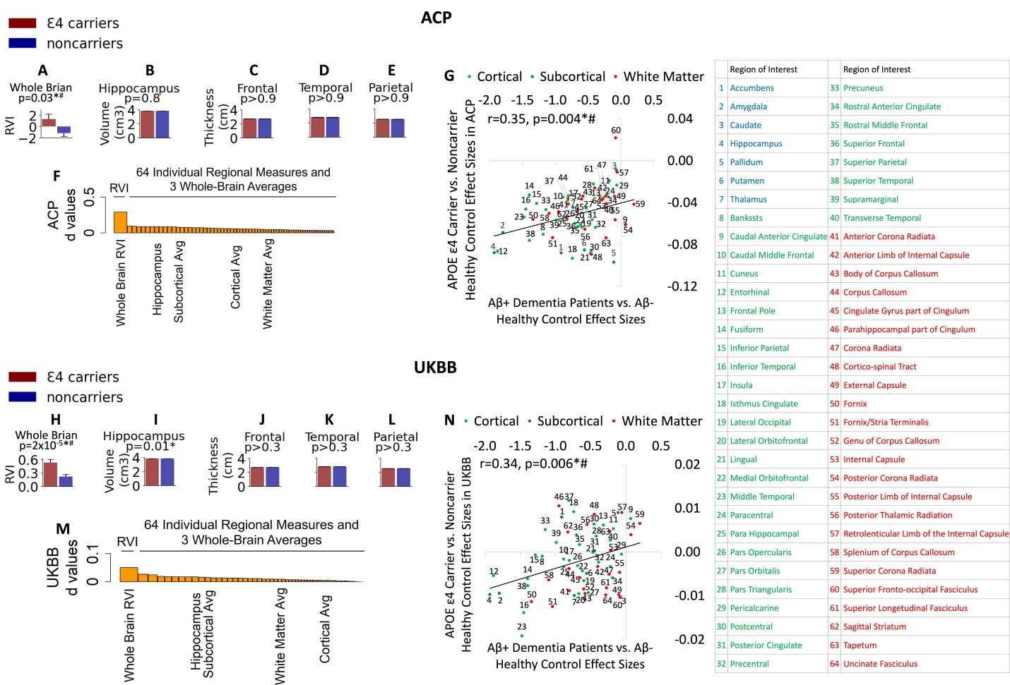
Preventing Alzheimer’s disease (AD) requires early-warning biomarkers. We developed a Regional Vulnerability Index (RVI) that quantifies individual brain similarity to AD patients' expected brain deficit patterns. We calculated regional effect sizes to establish brain deficit patterns in amyloid-positive AD cases compared to amyloid-negative healthy controls. The RVI-AD was calculated as a linear index of individual similarity to this established brain pattern in AD. Initially, we demonstrated RVI-AD elevation associated with risk factors in 335 participants (mean age: 49±13 years) in the Amish Connectome Project, followed by an independent sample consisting of 26,010 participants (mean age: 64±7 years) from the UK Biobank. Genetic and cardiovascular risks were evaluated using APOE-e4 genotype and Framingham Cardiovascular Risk Scores (FCVRS), respectively. Additionally, we assessed the risk of converting from MCI to dementia in N=1,932 participants (mean age: ~74) from the Alzheimer’s Disease Neuroimaging Initiative (ADNI). Participants with the APOE-e4 allele had significantly elevated RVI-AD indices (p<0.05); FCVRS significantly contributed to higher RVI-AD in an APOE-e4-specific manner (p<0.01), replicable across the samples. In the ADNI cohort, RVI-AD significantly predicted conversion from MCI to dementia in the next decade, particularly within the first 3 years (AUC=74%). In healthy individuals, the RVI-AD index detected the insidious impact of APOE-ε4 and cardiovascular risks in otherwise normally aging cohorts. Elevated RVI-AD also predicted conversion to dementia within 10 years in the older, high-risk cohort. Further development of this brain-pattern similarity-based approach may yield a noninvasive, clinically accessible biomarker to aid early detection of the subtle to more imminent effects of AD risks.
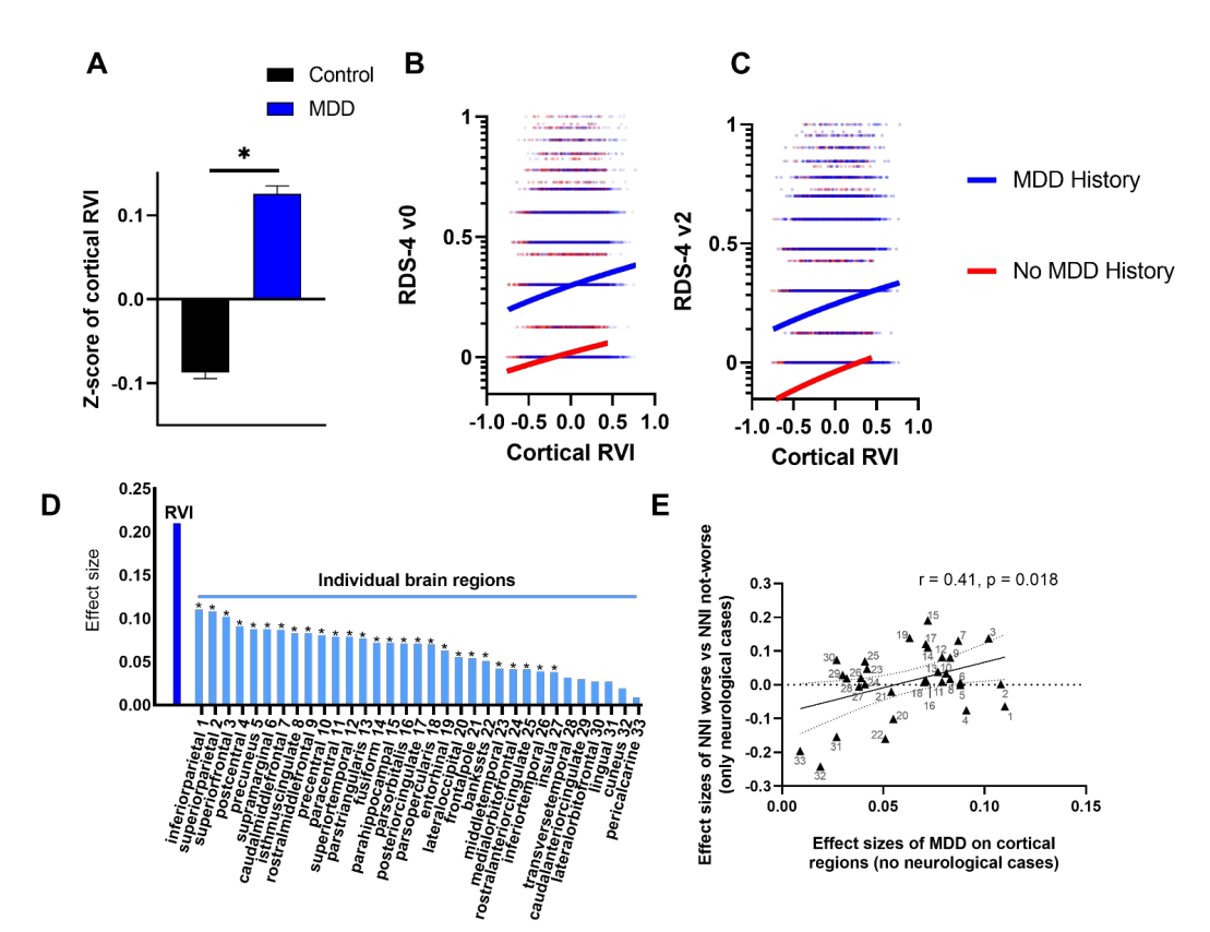
Depression Following Neurological Illnesses: Reshaping the Cortical Pattern Towards Major Depression
Mark D. Kvarta MD PhD, Yizhou Ma PhD, Si Gao MS, Jennifer Hopp MD, et al.
- Mark D. Kvarta MD, PhD, Yizhou Ma PhD, Si Gao MS, Jennifer Hopp MD, Bhim M. Adhikari PhD, Heather Bruce MD Joshua Chiappelli MD, Andrew van der Vaart MD, PhD, Xiaoming Du PhD, Paul M. Thompson, PhD, Jair Soares, MD, PhD, Carlos A. Zarate Jr. MD, Peter Kochunov PhD, L. Elliot Hong MD
Importance: In individuals with neurological illnesses, depression occurs in up to ~50% of cases, highlighting a common problem encountered on consultation-liaison psychiatry and neurology services. The neurobiological causes of this high rate of comorbid depressive disorders are unclear, and no valid biomarkers exist to guide prevention and treatment of depressive disorders due to neurological conditions. Objective: This study applied a Regional Vulnerability Index (RVI) to quantify individual brain-wide phenotypic similarity to primary major depressive disorder. The study sought to test the hypothesis that, after neurological insult, brain changes that result in structural similarity to depression would increase risk for having depressive symptoms. Design: The RVI was constructed using the cortical patterns of patient-control differences in depression reported by the largest independent meta-analysis to date to define brain cortical structural patterns of major depression. This cortical pattern was then applied to each UK Biobank sample participant to construct individual RVI. Setting: UK population. Participants: Individuals with new neurological incidents during the study period (n=890) and 46,020 non-neurological controls. Intervention: Longitudinal depressive symptoms were measured between the initial assessment visit (v0) and the second visit (v2), which took place about nine years apart. Main Outcome Measures: RVI in newly-reported major neurological illnesses during follow-up. Results: RVI significantly distinguished individuals with and without major depressive disorder history in participants who had no neurological illness (d=0.21, p=3.4x10-70). Significantly more participants in the group with new neurological conditions (30.8%) reported worsening depression scores between v0 and v2 than the non-neurological controls (24.9%, p=6x10⁻⁵). RVI showed a significant group*depression change interaction (F(1,35550)=4.02, p=0.045) wherein individuals who developed neurological conditions and reported worsened depression scores had the highest RVIs. Their RVIs were higher than those of non-neurological controls without (p=1x10⁻⁵) or with (p=0.001) worsening of depression in the same period, and higher than those of individuals who developed neurological conditions but did not report worsening of depression (p=0.01). Conclusion: Worsening depressive symptoms after new neurological events was associated with development of cortical patterns similar to major depression. RVI may provide a brain-pattern level biomarker to explain and capture vulnerability to developing depression associated with neurological illnesses.